


Process study / engineering - Food and agrifood technology
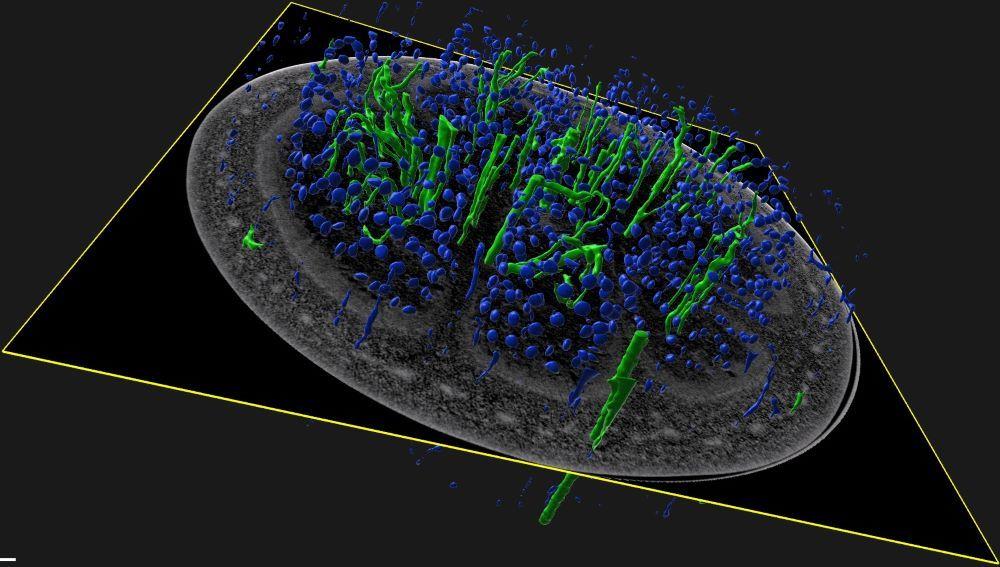
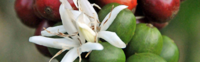
Eggplant observed in X-ray micro-tomography with segmentation of seeds (blue) and vascular tissues (green). © Marc Lartaud, CIRAD
LamHis technology enables the digitisation of histological sections mounted on glass slides. Beyond securing reference histological slides, it becomes possible to observe these digitised slides at very high resolution on a remote computer, just as with a microscope, and to explore the structure of plant organs at tissue, cellular and subcellular levels.
This method, already widely used in human and veterinary medicine, offers major potential for plant anatomy. LamHis provides access to this technology through dedicated tools and expert know-how made available by CIRAD.
This technique, adapted to plant science, enables:
LamHis is designed for a variety of professionals working with or teaching plant materials who wish to develop more advanced observation approaches: breeders, technologists, R&D engineers, slide curators, educational content developers and training institutions.
This service enables a wide range of applications for different stakeholders:
Preservation of slide collections and multi-user access to digital libraries.
Varietal selection and diagnosis; identification of areas and characteristics of interest in fibres; visualisation of extraction processes for compounds of interest and optimisation of extraction processes supported by imaging.
Illustration of courses and development of tutorials and practical sessions.
The service offering is structured around complementary services:
For more information on the plant histocytology and imaging platform : Click here
Process study / engineering - Food and agrifood technology
Software and digital applications - Territory management solution - Natural resources and territories
Process study / engineering - Food and agrifood technology - Food and agrifood technology