


- Home
- Work with us
- Open science
- CIRAD open infrastructures
- PHIV
Histocytology and plant cell imaging platform - PHIV
Experiments and analyses in a controlled environment
PHIV provides access to the latest techniques for visualizing the main molecules in the living world, both in situ and in vivo.
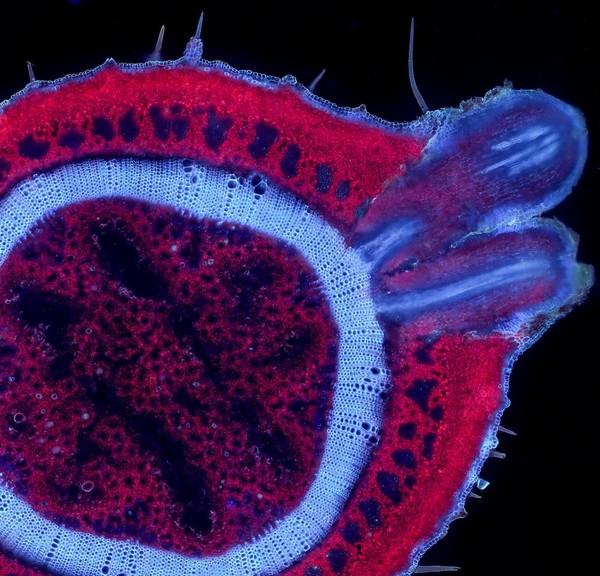
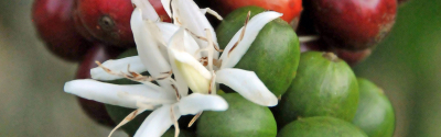
Jasmine cross-section © M. Lartaud, CIRAD-PHIV
The platform uses high-throughput imaging for plant quantitative genetics and design (anatomical phenotyping, elementary histochemistry). It also uses functional imaging to understand plant biology under stress (use of biosensors), from genes to organs and the whole plant.
PHIV works to interface 3D imaging and modelling, to facilitate the integration of knowledge obtained from functional genomics studies into plant development modelling programs, a vital issue for designing the plants of the future.